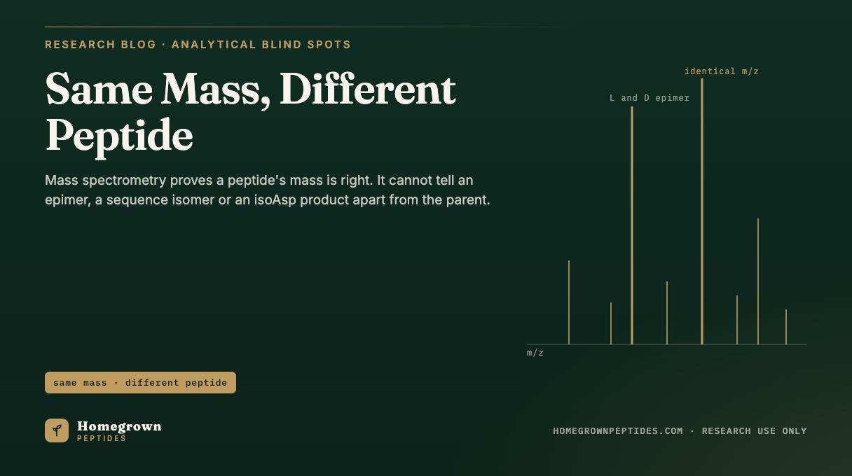
A mass spectrometer confirms that a molecule with the expected mass is present. It cannot, by itself, tell a peptide from its epimer (one residue flipped from L to D), its sequence isomer, or its isoaspartate rearrangement product, because all three have exactly the same mass. These isobaric impurities are real synthesis and storage products, and seeing them takes chromatography, MS/MS fragmentation, or chiral amino acid analysis.
| Impurity type | Mass difference from parent peptide | Seen by RP-HPLC? | Seen by standard MS? | Resolved by | Source |
|---|---|---|---|---|---|
| D-epimer (one residue inverted from L to D) | 0 Da, identical monoisotopic mass | Sometimes, as a close shoulder on the main peak | No, identical m/z and near-identical MS/MS fragment masses | Chiral amino acid analysis, differential ion mobility | Anal Chem, 2021 (PMID 33587599) |
| Sequence isomer (residue order transposed) | 0 Da | Often, hydrophobicity typically shifts more | Yes, MS/MS fragment ion masses differ by residue position | MS/MS sequencing (b and y ion ladders) | J Am Soc Mass Spectrom, 2019 (PMID 30421361) |
| IsoAsp (beta-linked aspartyl, from a succinimide intermediate) | 0 Da versus the normal alpha-linked Asp peptide | Often, tends to elute earlier as a shoulder | No by MS1 and no by routine CID; yes by electron-based fragmentation | ETD or ECD diagnostic backbone cleavages, enzymatic digests | Anal Chem, 2009 (PMID 19873993) |
| Leu/Ile substitution (isobaric side chains) | 0 Da | Rarely resolved on a generic gradient | No by standard collision-induced dissociation | Charge transfer dissociation or other radical-driven fragmentation | Rapid Commun Mass Spectrom, 2022 (PMID 34927767) |
What is an isobaric impurity?
An isobaric impurity has the same elemental formula and the same monoisotopic mass as the peptide it is contaminating, but a different shape or a different order of atoms along the chain. A mass spectrometer measures mass-to-charge ratio, so if two molecules weigh the same, it reports the same peak regardless of how the atoms are arranged. Mass Spectrometry for Peptide Identity covers how LC-MS proves a molecule's formula matches the intended target; this article covers the structures that pass that test without being the intended peptide.
There are three practical families. Stereoisomers, usually called epimers here, keep the same sequence and formula but have a residue in the wrong spatial configuration (D instead of the natural L form). Sequence isomers keep the same composition but place residues in a different order. Constitutional isomers change the backbone connectivity itself without changing the formula; isoaspartate and its succinimide precursor are the clearest peptide example.
Leucine and isoleucine are the textbook isobaric pair. Both have the formula C6H13NO2 and an identical residue mass near 113.084 Da; the only difference is where a methyl branch sits on the side chain. Ordinary tandem MS cannot tell them apart, because their fragment ions land at the same mass. A peptide built with an isoleucine where leucine belongs gives the same observed mass and, in most instruments, the same fragment ion table as the correct sequence.
How does epimerization happen during synthesis?
Most epimerization happens during carboxyl activation, not after the peptide is finished. When a residue's carboxyl group is activated for coupling, a five-membered oxazolone ring can close transiently next to its alpha carbon. That proton is unusually acidic, so a trace of base can pull it off and let it land back on either face of the ring, scrambling the stereocenter before the next coupling step locks it in place.
Histidine, cysteine, serine and phenylalanine show up repeatedly in the literature as the residues most prone to this, histidine in particular because its imidazole side chain can act as an internal base during its own activation. A second route runs through the base used to strip the Fmoc group every cycle; piperidine is mild, but a residue next to an electron-withdrawing group can still lose configuration under repeated exposure.
Coupling reagent and additive choice change how much of this happens. A 2014 study of aqueous, microwave-assisted Fmoc coupling through histidine-containing sequences measured racemization ranging from a small fraction of a percent up to several percent of crude product depending on the additive, with HOBt and Oxyma-type additives generally outperforming more basic conditions (PMID 24965528). Preparative HPLC removes some of what forms but not all, because a D-epimer can coelute closely with the parent peptide, covered further down. Solid-Phase Peptide Synthesis Explained covers the stepwise coupling chemistry itself; this section is only about where that cycle can flip a stereocenter.
What is aspartimide formation and why does it produce isoaspartate?
Aspartimide formation is a side reaction at aspartate and asparagine residues sitting next to a small, flexible residue, most often glycine or serine. Asp-Gly, Asp-Ser and Asn-Gly sequences are the recognized hot spots. Under the base used to remove Fmoc groups, the side-chain carbonyl of Asp or Asn can attack the backbone nitrogen of the following residue, closing a five-membered succinimide ring and releasing the side-chain protecting group or an ammonia equivalent.
That succinimide ring is an intermediate that reopens with water, and it can reopen at either of two carbonyls, which are not the same outcome. Reopening at the original position regenerates a normal alpha-linked aspartyl residue. Reopening at the other carbonyl produces a beta-linked isoaspartyl residue, isoAsp, in which the backbone now runs through what used to be the side chain. Both share the same formula and mass as the intended peptide, which is exactly why MS alone cannot flag the swap. The intermediate is also prone to racemization at its alpha carbon for the same oxazolone-adjacent reason above, so a single aspartimide event can hand back an isoAsp residue, a D-Asp residue, or both.
This is not purely a synthesis-time problem. The same chemistry runs during storage in aqueous solution near neutral pH, slowly converting correctly made Asp residues into isoAsp over time. How Peptides Degrade covers that storage-driven route alongside oxidation and hydrolysis. A 2003 study mapping aspartimide formation across model Fmoc sequences confirmed the Asp-Gly and Asp-Ser hot spots and how closely it tracks with base exposure (PMID 12952393).
Why can RP-HPLC sometimes separate them and sometimes not?
Reversed-phase HPLC separates molecules by how they interact with a hydrophobic stationary phase, which tracks overall hydrophobicity and folding, not strictly molecular formula. A D-epimer changes the local backbone geometry at one residue, shifting retention time measurably or barely at all depending on where it sits. In practice, D-epimers frequently elute close enough to the parent peptide that they show up only as a small shoulder, easy to fold into the main peak's integration.
IsoAsp-containing peptides behave more predictably: the beta-linked backbone usually elutes earlier than the alpha-linked parent on a standard gradient. Sequence isomers are the most likely of the group to separate cleanly, since swapping residue order can shift overall hydrophobicity more than one inverted stereocenter does. None of this is guaranteed either way; method, gradient and sequence all matter, which is why an HPLC purity number states what that particular method separated, not a universal claim about chiral or isomeric purity.
Which methods actually resolve isobaric impurities?
| Method | Distinguishes | Notes |
|---|---|---|
| MS/MS sequencing (CID or HCD) | Sequence isomers: yes. Epimers: no | Fragment ion masses move when residue order changes; they do not move when only stereochemistry changes |
| Ion mobility MS | Some epimers | Differential ion mobility has resolved D/L peptide epimers by their slightly different collision cross sections (PMID 33587599) |
| Chiral amino acid analysis after acid hydrolysis | Epimers (D versus L at specific residues) | Peptide is hydrolyzed to free amino acids, then derivatized (commonly with Marfey's reagent) or run on a chiral GC phase; hydrolysis in deuterated HCl is used to correct for racemization the hydrolysis step itself can introduce (PMID 30421361) |
| 2D-LC | Co-eluting epimers and isomers missed by one-dimensional RP-HPLC | A second, orthogonal separation mode catches what the first dimension cannot resolve |
| NMR | Structure and some stereochemistry | Requires more material and time than routine release testing allows |
| Edman degradation | Sequence isomers | Confirms residue order one position at a time; slow relative to MS/MS |
| Enzymatic digests | IsoAsp | Some proteases cleave differently around a beta-linked backbone than around the normal alpha linkage |
| Electron-based fragmentation (ETD, ECD) | IsoAsp specifically | Backbone N-Cα cleavage next to the isoAsp residue produces a diagnostic pair of fragment ions offset by about 57 Da from the corresponding Asp fragment pair, which routine CID does not generate (PMID 19873993, PMID 21472566) |
What does this mean for reading a certificate?
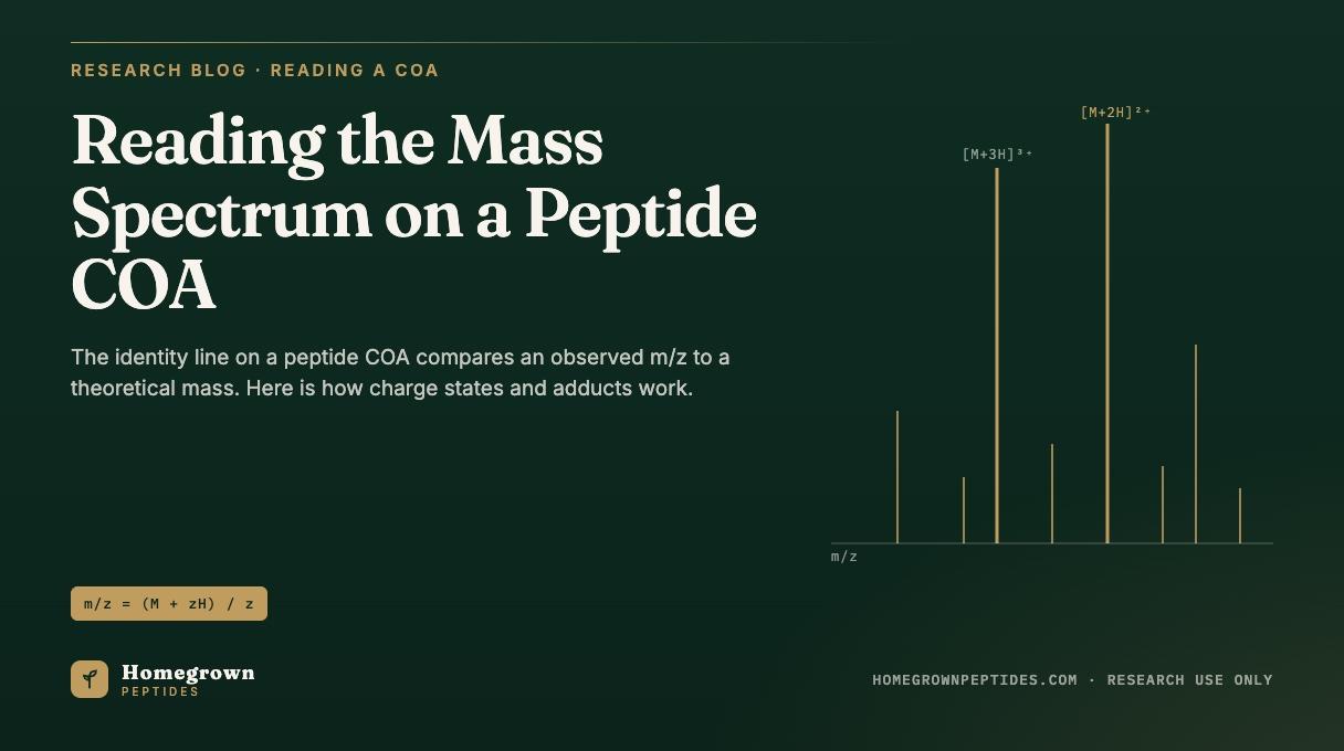
A purity percentage on a certificate of analysis reports what a specific HPLC method separated. An identity result from LC-MS, as Reading the Mass Spectrum on a Peptide COA explains for observed mass, charge states and adducts, reports that the dominant species in the vial has the expected mass within tolerance. Neither line claims chiral purity. A vial can report high HPLC purity and a clean identity match while still containing a measurable fraction of a D-epimer, a sequence isomer, or an isoAsp product, because none of those three routinely moves either number.
That gap reflects what each routine method is built to measure. A lab that specifically needs chiral purity information, for a certificate on a product such as PT-141 or any other synthetic peptide, can request chiral amino acid analysis for the residues most prone to racemization, documented MS/MS sequence coverage, or a written note on expected D-isomer content from the synthesis route used. Vendor certificates typically report intact mass, HPLC purity and sometimes net peptide content; they do not typically include chiral analysis unless specifically run, and silence on it should not be read as having ruled it out.
Do epimers matter for research results?
Published cell and animal studies on peptide analogs show that a single D-amino acid substitution can change receptor binding affinity and how quickly a proteolytic enzyme degrades the molecule, because binding pockets and protease active sites are themselves chiral. A 2016 survey of D-amino acid-containing neuropeptides built a workflow to find and characterize these naturally occurring and synthesis-derived epimers, on the premise that a single inverted residue measurably changes bioactivity in downstream assays (PMID 27788334). That is the practical reason an unintended epimer or isoAsp impurity is a quality concern and not only an analytical curiosity: the altered form can behave differently in the same binding or stability assay while reading identically on a routine LC-MS trace.
Frequently asked questions
Can LC-MS detect D-amino acids in a peptide?
Not on its own. A D-amino acid substitution does not change a residue's mass, so standard LC-MS, including MS/MS fragmentation, reports the same mass and typically the same fragment ion table for the D-epimer as for the correct L-form peptide. Detecting it requires a chirality-sensitive method such as chiral amino acid analysis or differential ion mobility.
What is an isobaric impurity?
It is a molecule with the same elemental formula and the same monoisotopic mass as the target peptide but a different structure: a flipped stereocenter, a transposed residue order, or an altered backbone connectivity such as isoAsp. Because the mass matches, a mass spectrometer alone cannot distinguish it from the correct peptide.
What is isoaspartate?
IsoAsp is a beta-linked form of aspartate in which the peptide backbone runs through the side-chain carbon rather than the normal alpha carbon. It forms through a succinimide (aspartimide) ring intermediate, most often at Asp-Gly, Asp-Ser or Asn-Gly sequences, either during synthesis under basic conditions or during storage near neutral pH.
Which residues racemize most during synthesis?
Histidine, cysteine, serine and phenylalanine are flagged most consistently in the synthesis literature, largely because their side chains or their local chemical environment make the oxazolone-mediated racemization pathway during carboxyl activation easier to reach.
Why does a certificate say identity confirmed if MS cannot see epimers?
"Identity confirmed" on a certificate of analysis means the observed mass matches the expected mass for the intended sequence within the method's tolerance. That is a real and useful result, but it is a statement about molecular formula, not about stereochemistry or backbone connectivity, so it does not rule out an epimer, a sequence isomer or an isoAsp product at the same mass.
How is chiral purity measured?
The peptide is hydrolyzed into its free amino acids, often using deuterated hydrochloric acid to correct for racemization the hydrolysis step itself can introduce, and the resulting amino acids are then separated and quantified by a chirality-sensitive method, commonly derivatization with Marfey's reagent followed by LC-MS/MS, or direct separation on a chiral gas chromatography phase.
References
- Mergler M, Dick F, Sax B, Stähelin C, Vorherr T. The aspartimide problem in Fmoc-based SPPS. Part II. J Pept Sci. 2003;9(8):518-526. PubMed PMID 12952393.
- Hojo K, Shinozaki N, Hidaka K, Tsuda Y, Fukumori Y, Ichikawa H, Wade JD. Aqueous microwave-assisted solid-phase peptide synthesis using Fmoc strategy. III: racemization studies and water-based synthesis of histidine-containing peptides. Amino Acids. 2014;46(10):2347-2354. PubMed PMID 24965528.
- Sargaeva NP, Lin C, O'Connor PB. Identification of aspartic and isoaspartic acid residues in amyloid beta peptides, including Abeta1-42, using electron-ion reactions. Anal Chem. 2009;81(23):9778-9786. PubMed PMID 19873993.
- Sargaeva NP, Lin C, O'Connor PB. Unusual fragmentation of β-linked peptides by ExD tandem mass spectrometry. J Am Soc Mass Spectrom. 2011;22(3):480-491. PubMed PMID 21472566.
- Berthias F, Baird MA, Shvartsburg AA. Differential ion mobility separations of d/l peptide epimers. Anal Chem. 2021;93(8):4015-4022. PubMed PMID 33587599.
- Ayon NJ, Sharma AD, Gutheil WG. LC-MS/MS-based separation and quantification of Marfey's reagent derivatized proteinogenic amino acid DL-stereoisomers. J Am Soc Mass Spectrom. 2019;30(3):448-458. PubMed PMID 30421361.
- Livnat I, Tai HC, Jansson ET, et al. A D-amino acid-containing neuropeptide discovery funnel. Anal Chem. 2016;88(23):11868-11876. PubMed PMID 27788334.
- Edwards HM, Wu HT, Julian RR, Jackson GP. Differentiation of leucine and isoleucine residues in peptides using charge transfer dissociation mass spectrometry (CTD-MS). Rapid Commun Mass Spectrom. 2022;36(5):e9246. PubMed PMID 34927767.
Research use only. Homegrown Peptides products are for laboratory research and are not for human or animal use. Nothing in this article is medical advice.