Almost everything on a peptide bench is a chain of amino acids. NAD+ is not. It is a dinucleotide coenzyme, and it behaves differently in storage, in solution and on an analytical column. If you are ordering it alongside peptides, the handling assumptions you carry over will be wrong in a few specific places. This article covers the chemistry and those places.
What the molecule is
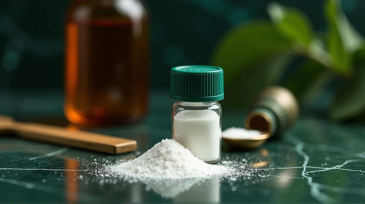
Nicotinamide adenine dinucleotide in its oxidized form is registered as CAS 53-84-9, with molecular formula C21H27N7O14P2 and a molecular weight of 663.43. It is also written as beta-NAD, and in older literature as coenzyme I. Structurally it is two nucleotides joined through their phosphate groups: one carrying adenine, the other carrying nicotinamide. There is no peptide bond anywhere in it. At 663 daltons it is also far smaller than most of the compounds around it, which is why it ships in 500 mg and 1000 mg vials rather than the 5 to 30 mg range typical of peptides.
The redox pair
The nicotinamide ring is the working end. It accepts a hydride ion to become NADH and gives it back to become NAD+ again. That two-electron shuttle is the central currency of catabolic metabolism, running through glycolysis, the tricarboxylic acid reactions and fatty acid oxidation on the oxidizing side and through the electron transport chain on the reducing side. In this role the coenzyme is a carrier: it is regenerated, not consumed, and the quantity in the cell stays constant while the ratio between the two forms moves.
That ratio is measurable, and it is the classic reason NAD+ appears in a laboratory in the first place. NADH absorbs strongly at 340 nanometres and NAD+ does not, so absorbance at 340 nm is the standard optical readout for any dehydrogenase assay. Both forms absorb at 260 nanometres through their adenine ring, which is the wavelength used for identity and concentration work on the purchased material.
The other job: a consumed substrate
The redox role is only half the story, and the half that gets less attention is the one driving most current research interest. Several enzyme families cleave NAD+ rather than recycling it, taking the ADP-ribose portion and releasing nicotinamide as a by-product.
- Sirtuins. A family of NAD+-dependent deacylases that remove acetyl and other acyl groups from lysine residues on histones and other proteins. Because they require NAD+ as a co-substrate, their activity is coupled to the cell's NAD+ supply rather than being independent of it.
- PARPs. Poly(ADP-ribose) polymerases build branched ADP-ribose polymers onto target proteins, most prominently in the DNA damage response. They turn over NAD+ rapidly when activated.
- CD38 and related glycohydrolases. These generate calcium-mobilizing second messengers from NAD+ and are another significant route of turnover.
Because these enzymes consume the coenzyme, the cell has to keep making it. That makes the supply side interesting in its own right.
The salvage pathway and NAMPT
Cells can build NAD+ from tryptophan de novo, but the dominant route in most mammalian tissues is the salvage pathway, which recycles the nicotinamide released by sirtuins and PARPs. The first and rate-limiting step is carried out by nicotinamide phosphoribosyltransferase, usually written NAMPT, which converts nicotinamide to nicotinamide mononucleotide. A second enzyme then adds the adenylyl group to complete the coenzyme. Revollo and colleagues established the link between this pathway and sirtuin activity in mammalian cells in 2004, and NAMPT has been a focus of the field ever since.
The published literature also reports declining NAD+ levels in various tissues with advancing age across several model organisms and tissue types. That observation, described in work by Verdin and others, is what connects the coenzyme to mitochondrial and cellular ageing research. It is a finding about tissue measurements in research models, not a statement about anything else.
Handling: where peptide habits go wrong
Three differences matter in practice.
- Light sensitivity in solution. The nicotinamide and adenine rings are chromophores, and reconstituted NAD+ degrades faster under light than most peptides do. Amber vials or foil, and a refrigerator with the door shut, are the standard answer.
- Heat and pH sensitivity. NAD+ hydrolyses at the glycosidic bond, and the rate climbs with temperature and with alkaline pH. Keep solutions cold, keep them near neutral or slightly acidic unless the assay says otherwise, and make them fresh where the assay allows.
- A different analytical method. HPLC of a nucleotide is not HPLC of a peptide. Detection is at 260 nm rather than 214 or 220 nm, and the chromatogram looks nothing like a peptide trace. Do not compare a NAD+ purity figure directly with a peptide's without checking the method, as explained in HPLC purity explained.
Mass is the other reason to slow down. A 1000 mg vial is roughly a hundred times the mass of a typical peptide vial, so reconstitution volumes and concentrations sit in a completely different range. Our storage and stability guide covers the general rules; take the light sensitivity note above as the NAD+-specific addition.
Formats and pairings we carry
Alongside the 500 mg and 1000 mg lyophilized vials, NAD+ is available as dissolving strips, a transmucosal format studied in delivery and formulation-comparison research rather than a vial to reconstitute. The NAD STACK bundle groups NAD+ with MOTS-c and SS-31, the two mitochondrial peptides most often run alongside it, each vial sealed with its own lot-matched certificate. Those three compounds and how they differ are compared in our mitochondrial research compounds guide.
Frequently asked questions
Is NAD+ a peptide?
No. It is a dinucleotide coenzyme with no amino acids and no peptide bonds. It is stocked alongside research peptides because it appears in the same experimental questions, not because it is chemically related to them.
What is the difference between NAD+ and NADH?
They are the oxidized and reduced forms of the same coenzyme. NAD+ accepts a hydride to become NADH, and NADH gives it up to become NAD+. The material supplied here is the oxidized form.
Why does the solution turn yellow?
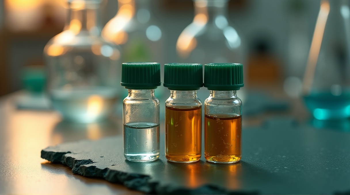
Yellowing usually indicates degradation, often from alkaline conditions, heat or prolonged light exposure. A fresh solution of the oxidized form should be essentially colourless. Discard discoloured solutions and note the lot.
References
- Cantó C, Menzies KJ, Auwerx J. 2015. NAD+ metabolism and the control of energy homeostasis. Cell Metabolism.
- Imai S, Guarente L. 2014. NAD+ and sirtuins in aging and disease. Trends in Cell Biology.
- Revollo JR, Grimm AA, Imai S. 2004. The NAD biosynthesis pathway mediated by nicotinamide phosphoribosyltransferase regulates Sir2 activity in mammalian cells. Journal of Biological Chemistry.
- Verdin E. 2015. NAD+ in aging, metabolism, and neurodegeneration. Science.
- Schreiber V, et al. 2006. Poly(ADP-ribose): novel functions for an old molecule. Nature Reviews Molecular Cell Biology.
Research use only. This article is analytical and biochemical background for laboratory researchers. Products in our research collection are not for human or animal use, are not drugs, and are not intended to diagnose, treat, cure or prevent any disease. Nothing here is medical advice.