Peptides aggregate when partially unfolded or locally hydrophobic segments on different molecules find each other faster than the molecules redissolve. The process is nucleation dependent: a slow lag phase builds a seed sized oligomer, and growth then accelerates into amorphous particles or ordered, cross beta amyloid like fibrils. Concentration, pH near the isoelectric point, ionic strength, agitation, air-water and container interfaces, and temperature all change the rate. Standard reversed-phase HPLC often misses the result entirely, which is why labs add size exclusion chromatography, light scattering, thioflavin T fluorescence and turbidity to the toolbox.
| Term | Definition | Detection method | Source |
|---|---|---|---|
| Nucleus | Smallest oligomer more likely to grow than dissolve; the rate limiting step. | Inferred from kinetic curves and seeding experiments. | Jarrett & Lansbury, 1993, PMID 8513491 |
| Lag phase | Period before detectable signal while nuclei form; minutes to weeks. | Time course ThT fluorescence or turbidity, 340 to 360 nm. | Knowles et al., 2014, PMID 24854788 |
| Secondary nucleation | New nuclei on existing fibril surfaces, not only at the ends. | Lag time with versus without added fibril seed. | Knowles et al., 2014, PMID 24854788 |
| Cross beta structure | Backbone arrangement in fibrils, strands roughly perpendicular to the axis. | ThT shift, Congo red birefringence, CD beta sheet signal. | Biancalana & Koide, 2010, PMID 20399286 |
| Thioflavin T (ThT) | Dye, weak free fluorescence, rises sharply once bound to fibril grooves. | Excitation about 440 nm, emission about 480 to 490 nm. | LeVine, 1999, PMID 10507030 |
| Hydrodynamic radius shift | Jump in apparent size once monomer assembles into oligomer or fibril. | Dynamic light scattering, about 1 nm to several microns. | Cromwell et al., 2006, PMID 17025275 |
| High molecular weight species (HMW) | Peak area eluting before the monomer peak, as percent of total. | SEC-HPLC, UV at 280 nm. | Cromwell et al., 2006, PMID 17025275 |
| Isoelectric point (pI) proximity | Net charge nears zero at the pI, removing repulsion between molecules. | Inferred from formulation pH versus calculated pI. | Chi et al., 2003, PMID 14567625 |
| Amyloid like fibril | Micrometer length filament of stacked cross beta sheets; resists mild redissolving. | TEM or AFM imaging, ThT positive. | Goldsbury et al., 2000, PMID 10940238 |
What is aggregation, physically?
Native, reversible self-association happens when correctly folded monomers stick together through normal surface contacts and can reverse on dilution. Non-native aggregation starts once a peptide partially unfolds or exposes a hydrophobic patch that is not normally solvent facing; once that patch meets a matching patch on another molecule, the result is usually much harder to reverse.
Amorphous aggregates are disordered clumps with no defined internal geometry; they scatter light and sometimes redissolve with heat, dilution or a pH change. Fibrillar aggregates have a cross beta architecture, extended beta strands stacked into sheets running the length of a long filament. That hydrogen bonded lattice makes amyloid like fibrils chemically stable and resistant to the mild conditions that clear up an amorphous aggregate. A short research peptide is not immune to either path; it only needs a stretch with high beta sheet propensity and enough hydrophobic character to make burying that surface against water favorable (Roberts, 2014, PMID 24908382; Chi et al., 2003, PMID 14567625).
How does nucleation-dependent polymerization work?
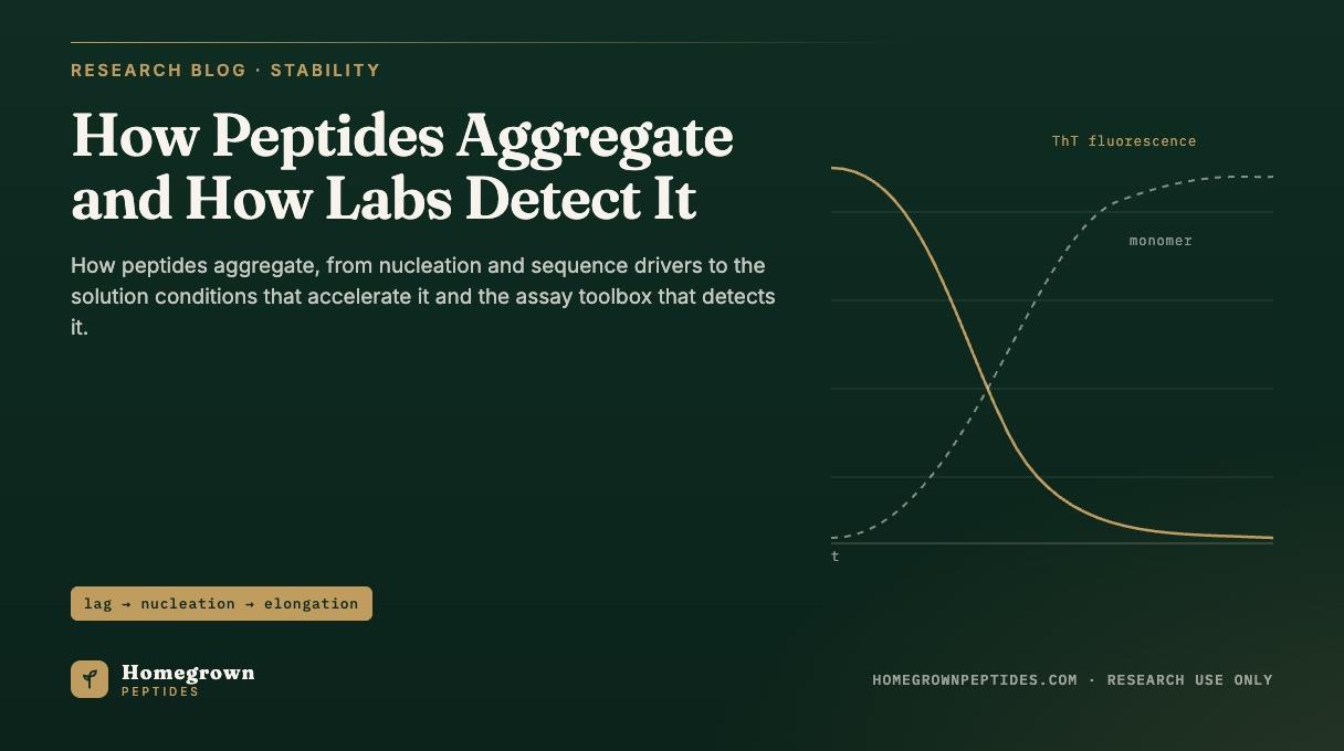
Peptide fibril formation generally follows nucleation dependent polymerization rather than a one-step reaction. A lone monomer is unstable as a growing filament; a small cluster arranged in the right geometry, the nucleus, is stable and tends to grow. Forming that nucleus by chance collision is slow, producing a lag phase of little detectable signal, followed by a rapid rise once nuclei exist and elongate. Plotted against time, the result is the sigmoidal curve seen in thioflavin T assays: flat, then a steep rise, then a plateau as monomer depletes (Jarrett & Lansbury, 1993, PMID 8513491).
Two features matter for a research lab. Seeding: adding a trace of preformed fibril to fresh monomer skips the slow nucleation step, shortening the lag phase sharply; a vial seeded by residue from glassware or a prior batch can behave very differently from a nominally identical fresh preparation. Secondary nucleation: once fibrils exist, their surfaces can catalyze new nuclei rather than only extending at the two ends, a major reason these reactions accelerate once underway (Knowles et al., 2014, PMID 24854788).
Which sequence features drive it?
Sequence sets the ceiling on aggregation risk. Long hydrophobic stretches, high intrinsic beta sheet propensity, a net charge close to neutral, and aromatic residues capable of stacking against each other all raise the odds that a given segment nucleates. Net charge matters because electrostatic repulsion between like charged molecules is one of the few forces that actively keeps peptides apart in solution (Roberts, 2014, PMID 24908382).
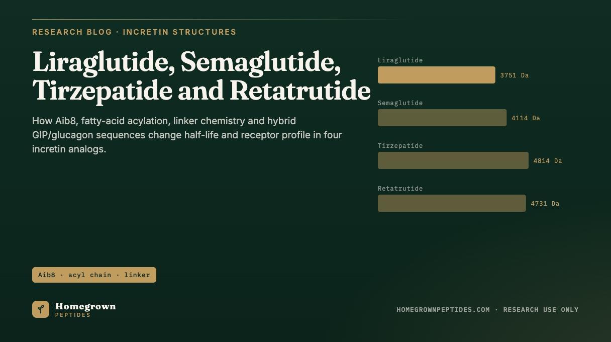
Chemical modification shifts this profile directly. Fatty acid acylation, the lipidation strategy used across the GLP-1 receptor agonist class described in the literature with codes such as NN9535, LY3298176 and LY3437943, adds a hydrophobic patch to extend plasma protein binding, the same kind of surface the aggregation literature flags as a self-association risk. That sequence logic is compared further in Liraglutide, NN9535, LY3298176 and LY3437943: How Sequence Modifications Change Half-Life and Receptor Profile, and carries over to amylin pathway analog research, including the AM833 class on the LY3437943 and AM833 certificate page. Amylin itself is one of the better studied amyloidogenic peptides outside the central nervous system literature (Goldsbury et al., 2000, PMID 10940238).
Which solution conditions accelerate it?
Sequence sets the ceiling; conditions decide how close a given preparation gets to it.
| Factor | Effect | Typical direction |
|---|---|---|
| Concentration | Rate scales faster than linearly with monomer concentration. | Higher, faster |
| pH relative to pI | Net charge is lowest near the pI, removing electrostatic repulsion. | Near pI, faster |
| Ionic strength | Salts can screen repulsion (Hofmeister type effect) or leave it intact, depending on the peptide. | Peptide specific |
| Temperature | Raises transient unfolding and collision frequency. | Higher, faster |
| Agitation / shear | Increases collisions and can locally unfold peptide at the point of highest stress. | More, faster |
| Air-water interface | More hydrophobic than bulk water; peptide partitions there, unfolds and concentrates. | Larger interface, faster |
| Container surfaces (glass, silicone) | Adsorption can locally unfold or concentrate peptide, seeding bulk aggregation. | More surface, faster |
| Metal ions (trace Fe, Cu) | Can catalyze oxidation of susceptible residues into a more aggregation prone species. | More trace metal, faster |
| Freeze-thaw cycling | Freeze concentration and new ice-water interfaces each cycle; a distinct mechanism. | Covered separately |
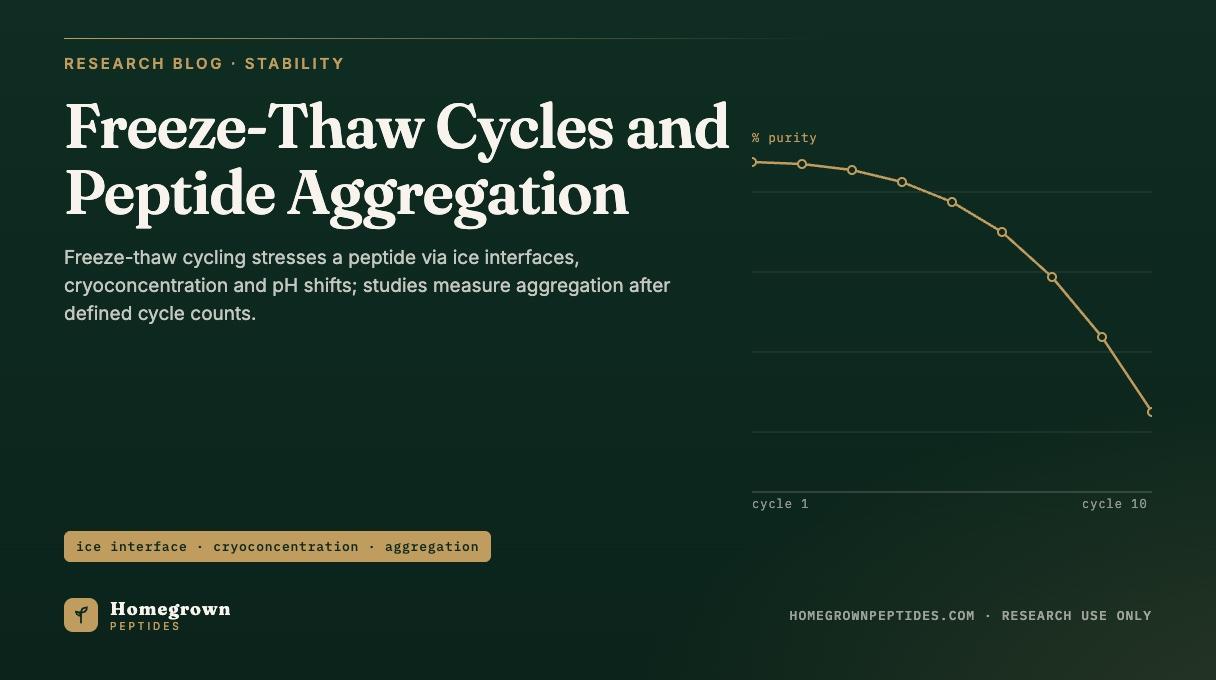
Freeze-thaw stress has its own kinetics and mitigation rules, covered in Freeze-Thaw Cycles and Peptide Aggregation: What the Stability Studies Show About Repeated Thawing and not repeated here. The remaining rows draw on a review of non-native aggregation driving forces (Chi et al., 2003, PMID 14567625) and on process level causes documented across peptide and protein bioprocessing (Cromwell et al., 2006, PMID 17025275). Solvent and buffer choice for reconstitution, which touches several of these rows at once, is covered in Peptide Solubility and Solvent Selection for Reconstitution.
Why does RP-HPLC miss aggregates?
Reversed-phase HPLC is the default purity method on most certificates, and it is largely blind to aggregation for three reasons. The mobile phase is denaturing: acetonitrile or methanol with trifluoroacetic acid at low pH dissociates most non-covalent aggregates into monomer before or during the run, so the chromatogram reports monomer purity regardless of what the vial contained. Larger aggregated species can adsorb irreversibly to the C18 stationary phase instead of eluting as a defined peak, removing them from the trace. Sample preparation also calls for a 0.2 or 0.22 micron syringe filter before injection, which strips visible and sub-visible particulate aggregates before the sample reaches the column.
The result is a method that can report a clean, high purity main peak from a solution that is visibly opalescent or would fail a light scattering test. Degradation pathways that run alongside aggregation, including oxidation and deamidation, are covered from the purity side in How Peptides Degrade: Oxidation, Deamidation, Hydrolysis and Aggregation (Cromwell et al., 2006, PMID 17025275).
Which assays see aggregates?
No single method spans soluble oligomer through visible particulate, so labs combine several orthogonal techniques.
| Method | What it detects | Size range | Strengths and limits |
|---|---|---|---|
| SEC-HPLC | Soluble oligomers and HMW species by size | About 1 to 100 nm | Quantitative, routine; column contact can dissociate weak aggregates |
| Dynamic light scattering (DLS) | Particle size distribution from scattered light | About 1 nm to several microns | Fast, no column; rare large particles can mask the main population |
| Static / multi-angle light scattering | Absolute molar mass, paired with SEC | About 10 nm to 1 micron | True molar mass, not apparent size |
| Thioflavin T fluorescence | Cross beta, fibrillar content | Fibrils only | Simple; negative result does not rule out amorphous aggregate |
| Congo red birefringence | Cross beta fibrils via polarized light | Fibrils only | Classic method; lower throughput than ThT |
| Turbidity (340 to 350 nm) | Bulk scattering from aggregate or precipitate | Submicron to visible | Very simple; not specific to aggregate identity |
| Micro-flow imaging | Sub-visible particle images, count and shape | About 1 to 100 microns | Separates particulate from bubbles or fibers by shape |
| Analytical ultracentrifugation | Sedimentation velocity across species | About 1 nm to 1 micron | High resolution reference method; slow |
| TEM / AFM imaging | Direct visualization of fibril morphology | Nanometer to micrometer | Unambiguous for fibrils; prep artifacts are a known pitfall |
| Native mass spectrometry | Oligomeric state, non-denaturing ionization | Monomer to small oligomers | Precise mass; large aggregates are harder to resolve |
Thioflavin T is the workhorse screen because its fluorescence turn on tracks the cross beta lattice rather than particle size generically, which is also why it misses amorphous aggregate (LeVine, 1999, PMID 10507030; Biancalana & Koide, 2010, PMID 20399286). SEC-HPLC and DLS are the two most common certificate entries because together they cover size ranges RP-HPLC alone cannot see (Cromwell et al., 2006, PMID 17025275).
What does aggregation look like on a certificate and in a vial?
Early amorphous aggregation often shows up as faint opalescence, a haze easy to miss under room light but visible against a dark background with a focused beam. More advanced aggregation produces visible particulates, fibers or a frank precipitate. A certificate line reading "appearance: clear, colorless solution" is a visual pass or fail check only; it confirms the absence of obvious haze at inspection, it does not quantify dissolved oligomer, and it says nothing about fibril content, since thioflavin T positive fibrils in dilute solution can be optically clear.
The more diagnostic entries are the SEC-HPLC high molecular weight percent, the fraction eluting ahead of the main monomer peak, and, on a stability program, the trend in main peak area over time. One SEC-HPLC result is a snapshot; a falling main peak area or a rising HMW percent across retest dates is the signal that aggregation, not a one-time handling error, is occurring in that lot.
Frequently asked questions
Can aggregated peptide redissolve?
Reversible self-association and many amorphous aggregates can redissolve with dilution, mild heating or a pH change away from the isoelectric point. Amyloid like fibrils, held by an extensive cross beta hydrogen bonding network, are far more resistant and generally do not redissolve under mild conditions.
Does lyophilized powder aggregate?
Aggregation needs molecular mobility, so it proceeds far more slowly in a dry cake than in solution. It is not impossible: residual moisture, an amorphous cake structure, or storage above the cake's glass transition temperature can allow slow solid state aggregation over long storage.
Is a cloudy solution always aggregation?
No. Cloudiness can also come from incomplete reconstitution, a precipitated excipient, microbial contamination or a container leachable. Aggregation is one differential diagnosis among several, distinguished by light scattering, filtration behavior and, when fibrils are suspected, a thioflavin T or Congo red result.
Does purity percent show aggregation?
Not reliably. A reversed-phase HPLC purity percentage is generated under denaturing mobile phase conditions that routinely dissociate non-covalent aggregates, so a high purity result and a real aggregate population can coexist in the same vial.
What is thioflavin T?
Thioflavin T is a small dye that fluoresces weakly in free solution and far more strongly once bound to the repeating grooves of a cross beta amyloid like fibril. The fluorescence increase, read on a standard plate reader, is the most widely used screening signal for fibril formation, though it does not detect amorphous aggregates.
Which peptides are most prone?
Prone sequences share features rather than a single identity: long hydrophobic stretches, high beta sheet propensity, a net charge near neutral at the formulation pH, and, for some engineered analogs, an added modification such as fatty acid acylation. Amylin and amylin pathway analogs are among the most studied amyloidogenic peptides in the literature; GLP-1 receptor agonist class analogs carrying lipidation, described with codes such as NN9535, LY3298176 and LY3437943, carry a related hydrophobic liability that formulation work is built around.
References
- Jarrett JT, Lansbury PT Jr. Seeding "one-dimensional crystallization" of amyloid: a pathogenic mechanism in Alzheimer's disease and scrapie? Cell. 1993;73(6):1055-1058. PubMed PMID 8513491.
- Knowles TPJ, Vendruscolo M, Dobson CM. The amyloid state and its association with protein misfolding diseases. Nat Rev Mol Cell Biol. 2014;15(6):384-396. PubMed PMID 24854788.
- Roberts CJ. Therapeutic protein aggregation: mechanisms, design, and control. Trends Biotechnol. 2014;32(7):372-380. PubMed PMID 24908382.
- Chi EY, Krishnan S, Randolph TW, Carpenter JF. Physical stability of proteins in aqueous solution: mechanism and driving forces in nonnative protein aggregation. Pharm Res. 2003;20(9):1325-1336. PubMed PMID 14567625.
- Cromwell MEM, Hilario E, Jacobson F. Protein aggregation and bioprocessing. AAPS J. 2006;8(3):E572-E579. PubMed PMID 17025275.
- LeVine H 3rd. Quantification of beta-sheet amyloid fibril structures with thioflavin T. Methods Enzymol. 1999;309:274-284. PubMed PMID 10507030.
- Biancalana M, Koide S. Molecular mechanism of Thioflavin-T binding to amyloid fibrils. Biochim Biophys Acta. 2010;1804(7):1405-1412. PubMed PMID 20399286.
- Goldsbury C, Goldie K, Pellaud J, et al. Amyloid fibril formation from full-length and fragments of amylin. J Struct Biol. 2000;130(2-3):352-362. PubMed PMID 10940238.
Research use only. Homegrown Peptides products are for laboratory research and are not for human or animal use. Nothing in this article is medical advice.